A new webinar calls attention to the importance of proper visual inspection of flexible endoscopes.
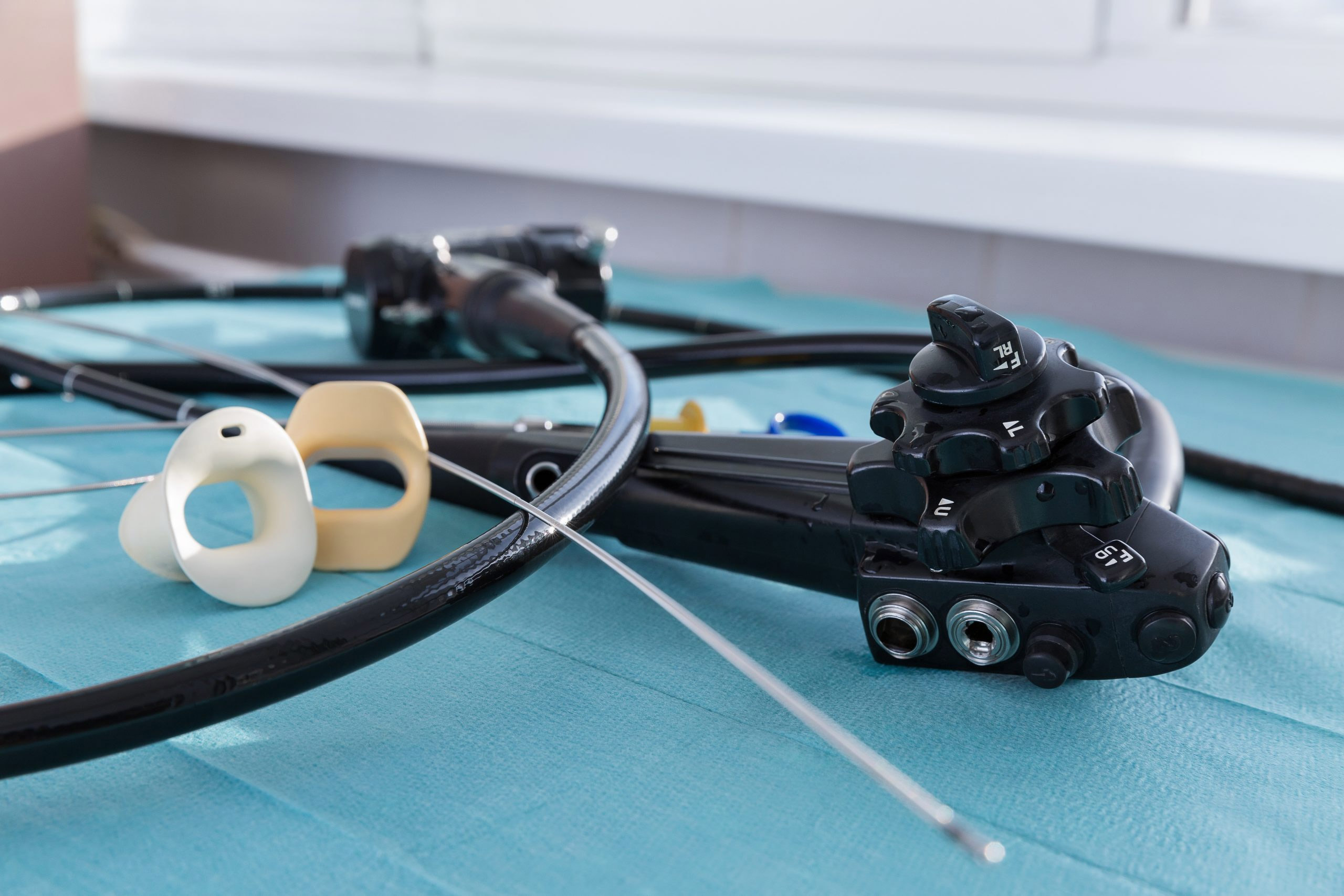
“Conducting visual inspections of flexible endoscopes using lighted magnification,” from epidemiologist Cori Ofstead, shines a light on the ways reusable endoscopes sustain various defects. Each type of endoscope features unique components that are prone to damage or residue collection.
Magnified and lighted visual inspection is a foundational technique essential to proper upkeep of a facility’s scopes. It should occur as part of reprocessing and prior to each use, she says.
Ofstead – with more than 25 years of research experience and whose firm works with a range of healthcare clients to improve the quality of medical care – shares photos she took of endoscopes displaying buckling, dents, crushing (dents on either side of the tube), scratches, and gouges. One contained patches of adhesive left behind by a poorly conducted repair job.
She also highlights injuries caused by damaged reusable gastroscopes, bronchoscopes, colonoscopes and ureteroscopes. A bronchoscope with a chipped plastic cover on the distal end, for example, cut a patient’s vocal cords. A lab later found fragments of the scope in tissue samples taken from the patient.
SGNA, AORN and AAMI guidelines recommend all reusable endoscopes be visually inspected, under good lighting and magnification, every time a scope is used.
Ofstead used a seemingly fine bronchoscope to underscore how important magnification is for visual inspections. An up-close look revealed brown residue packed along the sterilization cap.
Click here to register for the webinar. And click here for another webinar featuring Ofstead, on the best practices for bronchoscope use on COVID-19 patients.


A Learning Center by Ambu USA